Figure 1
A serious pulmonary infection secondary to disseminated Strongyloidiasis in a patient with Nephrotic syndrome
Mingming Ma, Shuang Cui, Ting Luo, Zhanhua Chen, Xiangnan Dong, Sibo Huang and Lianghong Yin*
Published: 03 April, 2019 | Volume 3 - Issue 1 | Pages: 071-075

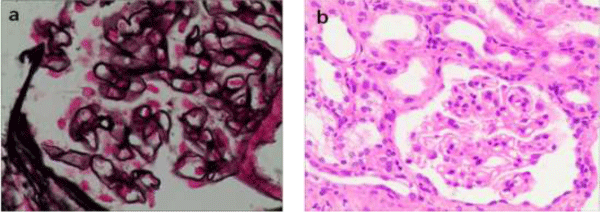
Figure 1:
1: (a) Light microscopy showing diffusely thickened basement membrane (Jones methenamine silver, original magnification x 400). (b) Light microscopy showing unremarkable proliferation of mesangial cell endothelial cells (hematoxylin and eosin, original magnification x200).
Read Full Article HTML DOI: 10.29328/journal.jcn.1001029 Cite this Article Read Full Article PDF
More Images
Similar Articles
-
A serious pulmonary infection secondary to disseminated Strongyloidiasis in a patient with Nephrotic syndromeMingming Ma,Shuang Cui,Ting Luo,Zhanhua Chen,Xiangnan Dong,Sibo Huang,Lianghong Yin*. A serious pulmonary infection secondary to disseminated Strongyloidiasis in a patient with Nephrotic syndrome. . 2019 doi: 10.29328/journal.jcn.1001029; 3: 071-075
-
A remote cause of anuria in a childMehtap Çelakıl*,Zelal Ekinci,Kürşat Demir Yıldız. A remote cause of anuria in a child. . 2020 doi: 10.29328/journal.jcn.1001050; 4: 011-013
-
Meanings of microalbuminuria in idiopathic nephrotic syndrome in childrenRichard Loumingou*. Meanings of microalbuminuria in idiopathic nephrotic syndrome in children. . 2020 doi: 10.29328/journal.jcn.1001051; 4: 014-015
-
Acute Kidney Injury due to spontaneous Atheroembolic disease, superimposed on diabetic nephropathy, with no recent vascular or cardiac intervention, presented as Rapidly Progressive Glomerulonephritis (RPGN)Anas Diab*,Parravani Anthony,Hollie Berryman,Kareem Diab. Acute Kidney Injury due to spontaneous Atheroembolic disease, superimposed on diabetic nephropathy, with no recent vascular or cardiac intervention, presented as Rapidly Progressive Glomerulonephritis (RPGN). . 2021 doi: 10.29328/journal.jcn.1001074; 5: 053-055
-
Practice patterns and outcomes of repository corticotropin injection (Acthar® Gel) use in childhood nephrotic syndrome: A study of the North American Pediatric Renal Trials and collaborative studies and the Pediatric Nephrology Research ConsortiumMohammed K Faizan*,Courtney McCracken,Kenneth Lieberman,Traci Leong,Mark R Benfield. Practice patterns and outcomes of repository corticotropin injection (Acthar® Gel) use in childhood nephrotic syndrome: A study of the North American Pediatric Renal Trials and collaborative studies and the Pediatric Nephrology Research Consortium. . 2021 doi: 10.29328/journal.jcn.1001077; 5: 067-076
-
Nephrotic syndrome in children during the COVID-19 pandemicAesha Maniar,Jenelle Cocorpus,Abby Basalely,Laura Castellanos,Pamela Singer,Christine B Sethna*. Nephrotic syndrome in children during the COVID-19 pandemic. . 2022 doi: 10.29328/journal.jcn.1001093; 6:
-
Strongyloides stercoralis and glomerular diseases: A case reportGabriel Ledesma*,Begoña Rivas,Cristina Vega,Gilda Carreño,Raquel Díaz,Ángel Gallegos,Verónica Mercado,Silvia Caldés,Yesika Amezquita,Yolanda Hernández,Rocío Echarri,Covadonga Hevia,Antonio Cirugeda. Strongyloides stercoralis and glomerular diseases: A case report. . 2022 doi: 10.29328/journal.jcn.1001099; 6: 097-098
-
Complications of ultrasound-guided percutaneous native kidney biopsies in children: A single center experienceAli H Asiri*,Musaed A AlQarni,Mohammed S Bafaqeeh,Abdulhadi M Altalhi,Abdulaziz A Alshathri,Khalid A Alsaran. Complications of ultrasound-guided percutaneous native kidney biopsies in children: A single center experience. . 2023 doi: 10.29328/journal.jcn.1001101; 7: 007-011
-
Mechanisms and Clinical Research Progress of Rituximab in the Treatment of Adult Minimal Change DiseaseYin Zheng, Hu Haofei, Wan Qijun*. Mechanisms and Clinical Research Progress of Rituximab in the Treatment of Adult Minimal Change Disease. . 2023 doi: 10.29328/journal.jcn.1001110; 7: 057-062
-
Doppler Evaluation of Renal Vessels in Pediatric Patients with Relapse and Remission in Different Categories of Nephrotic SyndromeAmit Nandan Dhar Dwivedi*, Srishti Sharma, OP Mishra, Girish Singh. Doppler Evaluation of Renal Vessels in Pediatric Patients with Relapse and Remission in Different Categories of Nephrotic Syndrome. . 2023 doi: 10.29328/journal.jcn.1001112; 7: 067-072
Recently Viewed
-
Various Theories of Fast and Ultrafast Magnetization DynamicsManfred Fähnle*. Various Theories of Fast and Ultrafast Magnetization Dynamics. Int J Phys Res Appl. 2024: doi: 10.29328/journal.ijpra.1001101; 7: 154-158
-
Forest History Association of WisconsinEd Bauer*. Forest History Association of Wisconsin. J Radiol Oncol. 2024: doi: 10.29328/journal.jro.1001071; 8: 093-096
-
Synthesis of Carbon Nano Fiber from Organic Waste and Activation of its Surface AreaHimanshu Narayan*,Brijesh Gaud,Amrita Singh,Sandesh Jaybhaye. Synthesis of Carbon Nano Fiber from Organic Waste and Activation of its Surface Area. Int J Phys Res Appl. 2019: doi: 10.29328/journal.ijpra.1001017; 2: 056-059
-
Obesity Surgery in SpainAniceto Baltasar*. Obesity Surgery in Spain. New Insights Obes Gene Beyond. 2020: doi: 10.29328/journal.niogb.1001013; 4: 013-021
-
Tamsulosin and Dementia in old age: Is there any relationship?Irami Araújo-Filho*,Rebecca Renata Lapenda do Monte,Karina de Andrade Vidal Costa,Amália Cinthia Meneses Rêgo. Tamsulosin and Dementia in old age: Is there any relationship?. J Neurosci Neurol Disord. 2019: doi: 10.29328/journal.jnnd.1001025; 3: 145-147
Most Viewed
-
Evaluation of Biostimulants Based on Recovered Protein Hydrolysates from Animal By-products as Plant Growth EnhancersH Pérez-Aguilar*, M Lacruz-Asaro, F Arán-Ais. Evaluation of Biostimulants Based on Recovered Protein Hydrolysates from Animal By-products as Plant Growth Enhancers. J Plant Sci Phytopathol. 2023 doi: 10.29328/journal.jpsp.1001104; 7: 042-047
-
Sinonasal Myxoma Extending into the Orbit in a 4-Year Old: A Case PresentationJulian A Purrinos*, Ramzi Younis. Sinonasal Myxoma Extending into the Orbit in a 4-Year Old: A Case Presentation. Arch Case Rep. 2024 doi: 10.29328/journal.acr.1001099; 8: 075-077
-
Feasibility study of magnetic sensing for detecting single-neuron action potentialsDenis Tonini,Kai Wu,Renata Saha,Jian-Ping Wang*. Feasibility study of magnetic sensing for detecting single-neuron action potentials. Ann Biomed Sci Eng. 2022 doi: 10.29328/journal.abse.1001018; 6: 019-029
-
Pediatric Dysgerminoma: Unveiling a Rare Ovarian TumorFaten Limaiem*, Khalil Saffar, Ahmed Halouani. Pediatric Dysgerminoma: Unveiling a Rare Ovarian Tumor. Arch Case Rep. 2024 doi: 10.29328/journal.acr.1001087; 8: 010-013
-
Physical activity can change the physiological and psychological circumstances during COVID-19 pandemic: A narrative reviewKhashayar Maroufi*. Physical activity can change the physiological and psychological circumstances during COVID-19 pandemic: A narrative review. J Sports Med Ther. 2021 doi: 10.29328/journal.jsmt.1001051; 6: 001-007

HSPI: We're glad you're here. Please click "create a new Query" if you are a new visitor to our website and need further information from us.
If you are already a member of our network and need to keep track of any developments regarding a question you have already submitted, click "take me to my Query."


