Figure 2
Membranous nephropathy complicating relapsing polychondritis: A case report
Christopher Rice, Vatsalya Kosuru, John Jason White, Christine Van Beek, Rachel Elam*, Michael Clemenshaw, Laura Carbone and Leighton James
Published: 07 October, 2021 | Volume 5 - Issue 3 | Pages: 084-087

Figure 2:
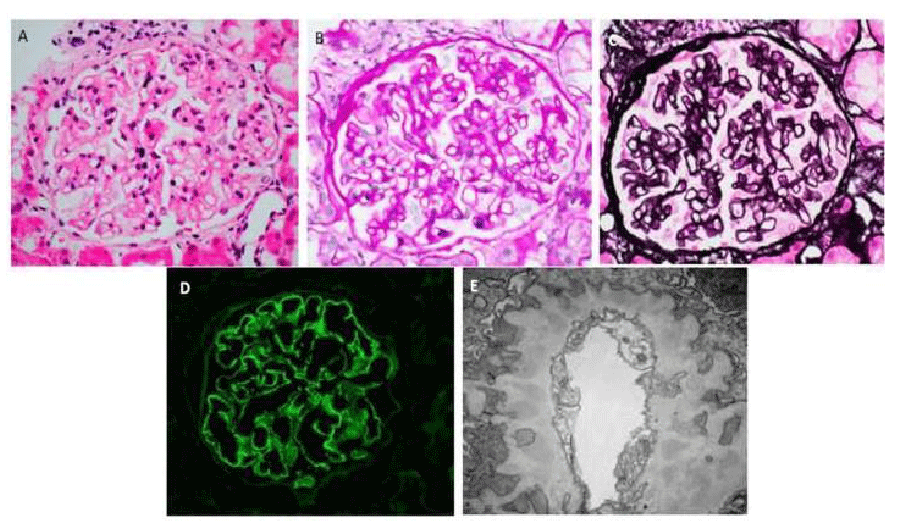
Kidney Biopsy Demonstrating Membranous Nephropathy. A. Hematoxylin and eosin stain (original magnification x400): Normocellular glomerulus. B. Periodic acid-Schiff stain (original magnification x400): Image shows a glomerulus with slight mesangial prominence and irregular capillary wall thickening. There is no significant hypercellularity. C. Jones methenamine silver stain (original magnification x400): Holes and occasional spikes are visible along glomerular capillary basement membranes. D. IgG immunofluorescence (original magnification x400): IgG is positive in glomerular capillary walls in a granular pattern. E. Electron microscopy (original magnification x20,000): There are continuous subepithelial deposits along glomerular capillary walls, which are well-incorporated into basement membranes and separated or occasionally surrounded by projections of basement membrane material consistent with Churg and Ehrenreich ultrastructural stage II to III membranous nephropathy [8].
Read Full Article HTML DOI: 10.29328/journal.jcn.1001080 Cite this Article Read Full Article PDF
More Images
Similar Articles
-
Acute Tubulointerstitial Nephritis due to Phenytoin: Case ReportNilzete Liberato Bresolin*,Pedro Docusse Junior,Maria Beatriz Cacese Shiozawa,Marina Ratier de Brito Moreira,Natalia Galbiatti Silveira. Acute Tubulointerstitial Nephritis due to Phenytoin: Case Report. . 2017 doi: 10.29328/journal.jcn.1001004; 1: 019-025
-
Equine Anti-Thymocyte Globulin (ATGAM) administration in patient with previous rabbit Anti-Thymocyte Globulin (Thymoglobulin) induced serum sickness: A case reportJoseph B Pryor*,Ali J Olyaei,Joseph B Lockridge,Douglas J Norman. Equine Anti-Thymocyte Globulin (ATGAM) administration in patient with previous rabbit Anti-Thymocyte Globulin (Thymoglobulin) induced serum sickness: A case report. . 2018 doi: 10.29328/journal.jcn.1001013; 2: 015-019
-
A case report of Hypocomplementemic urticarial vasculitic syndrome presenting with Renal failureAmy M Hopkins,Angela M Gibbs,Ryan S Griffiths,Rupali S Avasare,Firas G Khoury*. A case report of Hypocomplementemic urticarial vasculitic syndrome presenting with Renal failure. . 2018 doi: 10.29328/journal.jcn.1001017; 2: 039-043
-
Lessons from the success and failures of peritoneal Dialysis-Related Brucella Peritonitis in the last 16 years: Case report and Literature reviewMuhammad Bukhari*,Azharuddin Mohammed,Abdullah Al-Guraibi,Ahmed Shahata,Mansour Al-Ghamdi. Lessons from the success and failures of peritoneal Dialysis-Related Brucella Peritonitis in the last 16 years: Case report and Literature review. . 2018 doi: 10.29328/journal.jcn.1001020; 2: 057-061
-
Anti-glomerular basement membrane disease: A case report of an uncommon presentationNatália Silva*,Luís Oliveira,Mónica Frutuoso,Teresa Morgado. Anti-glomerular basement membrane disease: A case report of an uncommon presentation. . 2019 doi: 10.29328/journal.jcn.1001027; 3: 061-065
-
Granulomatosis with polyangiitis (GPA) in a 76-year old woman presenting with pulmonary nodule and accelerating acute kidney injuryNoor Sameh Darwich*,Melissa Schnell,L. Nicholas Cossey. Granulomatosis with polyangiitis (GPA) in a 76-year old woman presenting with pulmonary nodule and accelerating acute kidney injury. . 2020 doi: 10.29328/journal.jcn.1001048; 4: 001-006
-
Unusual presentation of oxalate nephropathy causing acute kidney injury: A case reportAnas Diab*,Michelle M Neuman,Kareem Diab,Daniel Gordon. Unusual presentation of oxalate nephropathy causing acute kidney injury: A case report. . 2020 doi: 10.29328/journal.jcn.1001063; 4: 077-079
-
Prostate cancer-associated thrombotic microangiopathy: A case report and review of the literatureHHS Kharagjitsing*,PAW te Boekhorst,Nazik Durdu-Rayman. Prostate cancer-associated thrombotic microangiopathy: A case report and review of the literature. . 2021 doi: 10.29328/journal.jcn.1001068; 5: 017-022
-
Membranous nephropathy complicating relapsing polychondritis: A case reportChristopher Rice,Vatsalya Kosuru,John Jason White,Christine Van Beek,Rachel Elam*,Michael Clemenshaw,Laura Carbone,Leighton James. Membranous nephropathy complicating relapsing polychondritis: A case report. . 2021 doi: 10.29328/journal.jcn.1001080; 5: 084-087
-
Persistent symptomatic hyponatremia post-COVID 19: case reportHaifa Alshwikh,Ferial Alshwikh,Halla Elshwekh*. Persistent symptomatic hyponatremia post-COVID 19: case report. . 2022 doi: 10.29328/journal.jcn.1001090; 6: 058-062
Recently Viewed
-
Various Theories of Fast and Ultrafast Magnetization DynamicsManfred Fähnle*. Various Theories of Fast and Ultrafast Magnetization Dynamics. Int J Phys Res Appl. 2024: doi: 10.29328/journal.ijpra.1001101; 7: 154-158
-
Forest History Association of WisconsinEd Bauer*. Forest History Association of Wisconsin. J Radiol Oncol. 2024: doi: 10.29328/journal.jro.1001071; 8: 093-096
-
Synthesis of Carbon Nano Fiber from Organic Waste and Activation of its Surface AreaHimanshu Narayan*,Brijesh Gaud,Amrita Singh,Sandesh Jaybhaye. Synthesis of Carbon Nano Fiber from Organic Waste and Activation of its Surface Area. Int J Phys Res Appl. 2019: doi: 10.29328/journal.ijpra.1001017; 2: 056-059
-
Obesity Surgery in SpainAniceto Baltasar*. Obesity Surgery in Spain. New Insights Obes Gene Beyond. 2020: doi: 10.29328/journal.niogb.1001013; 4: 013-021
-
Tamsulosin and Dementia in old age: Is there any relationship?Irami Araújo-Filho*,Rebecca Renata Lapenda do Monte,Karina de Andrade Vidal Costa,Amália Cinthia Meneses Rêgo. Tamsulosin and Dementia in old age: Is there any relationship?. J Neurosci Neurol Disord. 2019: doi: 10.29328/journal.jnnd.1001025; 3: 145-147
Most Viewed
-
Evaluation of Biostimulants Based on Recovered Protein Hydrolysates from Animal By-products as Plant Growth EnhancersH Pérez-Aguilar*, M Lacruz-Asaro, F Arán-Ais. Evaluation of Biostimulants Based on Recovered Protein Hydrolysates from Animal By-products as Plant Growth Enhancers. J Plant Sci Phytopathol. 2023 doi: 10.29328/journal.jpsp.1001104; 7: 042-047
-
Sinonasal Myxoma Extending into the Orbit in a 4-Year Old: A Case PresentationJulian A Purrinos*, Ramzi Younis. Sinonasal Myxoma Extending into the Orbit in a 4-Year Old: A Case Presentation. Arch Case Rep. 2024 doi: 10.29328/journal.acr.1001099; 8: 075-077
-
Feasibility study of magnetic sensing for detecting single-neuron action potentialsDenis Tonini,Kai Wu,Renata Saha,Jian-Ping Wang*. Feasibility study of magnetic sensing for detecting single-neuron action potentials. Ann Biomed Sci Eng. 2022 doi: 10.29328/journal.abse.1001018; 6: 019-029
-
Pediatric Dysgerminoma: Unveiling a Rare Ovarian TumorFaten Limaiem*, Khalil Saffar, Ahmed Halouani. Pediatric Dysgerminoma: Unveiling a Rare Ovarian Tumor. Arch Case Rep. 2024 doi: 10.29328/journal.acr.1001087; 8: 010-013
-
Physical activity can change the physiological and psychological circumstances during COVID-19 pandemic: A narrative reviewKhashayar Maroufi*. Physical activity can change the physiological and psychological circumstances during COVID-19 pandemic: A narrative review. J Sports Med Ther. 2021 doi: 10.29328/journal.jsmt.1001051; 6: 001-007

HSPI: We're glad you're here. Please click "create a new Query" if you are a new visitor to our website and need further information from us.
If you are already a member of our network and need to keep track of any developments regarding a question you have already submitted, click "take me to my Query."


