Figure 1
Kidney Biopsy in Autosomal Dominant Polycystic Kidney Disease
Laalasa Varanasi*, Gabriel Loeb, Vighnesh Walavalkar, Nebil Mohammed, John Paul Lindsey II, Stephen Gluck, Thomas Lee Chi and Meyeon Park
Published: 19 December, 2023 | Volume 7 - Issue 3 | Pages: 101-105

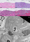
Figure 1:
Figure 1: a) Light microscopy with various stains (clockwise starting at top left: hematoxylin & eosin, periodic acid Schiff, Masson’s trichrome, Jones methenamine silver) showing the perihilar variant of FSGS. The circled area highlights the consolidation of capillary loops with hyaline deposition and intracapillary foam cells.
b) Electron microscopy showing moderate, not diffuse, effacement of podocyte foot processes suggestive of secondary FSGS.
Read Full Article HTML DOI: 10.29328/journal.jcn.1001118 Cite this Article Read Full Article PDF
More Images
Similar Articles
-
Liver cyst infection in kidney transplant patient with autosomal dominant polycystic kidney disease: Interest of PET/CT in diagnosis and treatmentGeorgery H*,Migali G,Pochet JM,Tintillier M,Van Ende C,Cuvelier C. Liver cyst infection in kidney transplant patient with autosomal dominant polycystic kidney disease: Interest of PET/CT in diagnosis and treatment. . 2018 doi: 10.29328/journal.jcn.1001019; 2: 053-056
-
Hypocomplementemic interstitial nephritis with long-term follow-upAlyssa Penning, M.D,Claire Kassakian, M.D,Donald C Houghton, M.D,Nicole K Andeen, M.D*. Hypocomplementemic interstitial nephritis with long-term follow-up. . 2019 doi: 10.29328/journal.jcn.1001024; 3: 042-045
-
Anti-glomerular basement membrane disease: A case report of an uncommon presentationNatália Silva*,Luís Oliveira,Mónica Frutuoso,Teresa Morgado. Anti-glomerular basement membrane disease: A case report of an uncommon presentation. . 2019 doi: 10.29328/journal.jcn.1001027; 3: 061-065
-
Cytomegalovirus infection in native kidney biopsyRaquel M Moreira,Géssica SB Barbosa*,Cristiane B Dias,Luis Yu,Viktoria Woronik,Lívia B Cavalcante. Cytomegalovirus infection in native kidney biopsy. . 2019 doi: 10.29328/journal.jcn.1001047; 3: 186-186
-
Granulomatosis with polyangiitis (GPA) in a 76-year old woman presenting with pulmonary nodule and accelerating acute kidney injuryNoor Sameh Darwich*,Melissa Schnell,L. Nicholas Cossey. Granulomatosis with polyangiitis (GPA) in a 76-year old woman presenting with pulmonary nodule and accelerating acute kidney injury. . 2020 doi: 10.29328/journal.jcn.1001048; 4: 001-006
-
A remote cause of anuria in a childMehtap Çelakıl*,Zelal Ekinci,Kürşat Demir Yıldız. A remote cause of anuria in a child. . 2020 doi: 10.29328/journal.jcn.1001050; 4: 011-013
-
Acute Kidney Injury due to spontaneous Atheroembolic disease, superimposed on diabetic nephropathy, with no recent vascular or cardiac intervention, presented as Rapidly Progressive Glomerulonephritis (RPGN)Anas Diab*,Parravani Anthony,Hollie Berryman,Kareem Diab. Acute Kidney Injury due to spontaneous Atheroembolic disease, superimposed on diabetic nephropathy, with no recent vascular or cardiac intervention, presented as Rapidly Progressive Glomerulonephritis (RPGN). . 2021 doi: 10.29328/journal.jcn.1001074; 5: 053-055
-
Membranous nephropathy complicating relapsing polychondritis: A case reportChristopher Rice,Vatsalya Kosuru,John Jason White,Christine Van Beek,Rachel Elam*,Michael Clemenshaw,Laura Carbone,Leighton James. Membranous nephropathy complicating relapsing polychondritis: A case report. . 2021 doi: 10.29328/journal.jcn.1001080; 5: 084-087
-
Administration of G-CSF for PBSC collection may unmask pre-existing IgA-nephropathy: A case reportFlorian Obereisenbuchner*,Sabine Bader-Zollner,Hans-Paul Schobel. Administration of G-CSF for PBSC collection may unmask pre-existing IgA-nephropathy: A case report. . 2022 doi: 10.29328/journal.jcn.1001094; 6: 079-082
-
Evaluation of the relationship between serum uric acid level and proteinuria in patients with type 2 diabetesMehrdad Chalak,Mehran Farajollahi,Majid Dastorani,Saeid Amirkhanlou*. Evaluation of the relationship between serum uric acid level and proteinuria in patients with type 2 diabetes. . 2023 doi: 10.29328/journal.jcn.1001100; 7: 001-006
Recently Viewed
-
Sensitivity and Intertextile variance of amylase paper for saliva detectionAlexander Lotozynski*. Sensitivity and Intertextile variance of amylase paper for saliva detection. J Forensic Sci Res. 2020: doi: 10.29328/journal.jfsr.1001017; 4: 001-003
-
Extraction of DNA from face mask recovered from a kidnapping sceneBassey Nsor*,Inuwa HM. Extraction of DNA from face mask recovered from a kidnapping scene. J Forensic Sci Res. 2022: doi: 10.29328/journal.jfsr.1001029; 6: 001-005
-
The Ketogenic Diet: The Ke(y) - to Success? A Review of Weight Loss, Lipids, and Cardiovascular RiskAngela H Boal*, Christina Kanonidou. The Ketogenic Diet: The Ke(y) - to Success? A Review of Weight Loss, Lipids, and Cardiovascular Risk. J Cardiol Cardiovasc Med. 2024: doi: 10.29328/journal.jccm.1001178; 9: 052-057
-
Could apple cider vinegar be used for health improvement and weight loss?Alexander V Sirotkin*. Could apple cider vinegar be used for health improvement and weight loss?. New Insights Obes Gene Beyond. 2021: doi: 10.29328/journal.niogb.1001016; 5: 014-016
-
Maximizing the Potential of Ketogenic Dieting as a Potent, Safe, Easy-to-Apply and Cost-Effective Anti-Cancer TherapySimeon Ikechukwu Egba*,Daniel Chigbo. Maximizing the Potential of Ketogenic Dieting as a Potent, Safe, Easy-to-Apply and Cost-Effective Anti-Cancer Therapy. Arch Cancer Sci Ther. 2025: doi: 10.29328/journal.acst.1001047; 9: 001-005
Most Viewed
-
Evaluation of Biostimulants Based on Recovered Protein Hydrolysates from Animal By-products as Plant Growth EnhancersH Pérez-Aguilar*, M Lacruz-Asaro, F Arán-Ais. Evaluation of Biostimulants Based on Recovered Protein Hydrolysates from Animal By-products as Plant Growth Enhancers. J Plant Sci Phytopathol. 2023 doi: 10.29328/journal.jpsp.1001104; 7: 042-047
-
Sinonasal Myxoma Extending into the Orbit in a 4-Year Old: A Case PresentationJulian A Purrinos*, Ramzi Younis. Sinonasal Myxoma Extending into the Orbit in a 4-Year Old: A Case Presentation. Arch Case Rep. 2024 doi: 10.29328/journal.acr.1001099; 8: 075-077
-
Feasibility study of magnetic sensing for detecting single-neuron action potentialsDenis Tonini,Kai Wu,Renata Saha,Jian-Ping Wang*. Feasibility study of magnetic sensing for detecting single-neuron action potentials. Ann Biomed Sci Eng. 2022 doi: 10.29328/journal.abse.1001018; 6: 019-029
-
Pediatric Dysgerminoma: Unveiling a Rare Ovarian TumorFaten Limaiem*, Khalil Saffar, Ahmed Halouani. Pediatric Dysgerminoma: Unveiling a Rare Ovarian Tumor. Arch Case Rep. 2024 doi: 10.29328/journal.acr.1001087; 8: 010-013
-
Physical activity can change the physiological and psychological circumstances during COVID-19 pandemic: A narrative reviewKhashayar Maroufi*. Physical activity can change the physiological and psychological circumstances during COVID-19 pandemic: A narrative review. J Sports Med Ther. 2021 doi: 10.29328/journal.jsmt.1001051; 6: 001-007

HSPI: We're glad you're here. Please click "create a new Query" if you are a new visitor to our website and need further information from us.
If you are already a member of our network and need to keep track of any developments regarding a question you have already submitted, click "take me to my Query."


